Case Study
Increasing imaging quality using Machine Learning


Author



Professor Carola Bibiane Schönlieb
Keywords
image analysis
partial differential equations
neural networks
Overview
The Project
Combining the best of both worlds
In a push towards cheaper, less harmful, technologies, Dr Ander Biguri’s Computational Applied Tomography (CAT) group and CHIMiRA mathematician Carola-Bibiane Schönlieb are working closely with radiologists on the development of algorithms. Their goal is to enable faster scans, that would expose patients to less radiation but would still be able to act as an accurate diagnosis tool. Building on 15 years’ experience of image reconstruction, they are combining the accuracy and capability of neural networks with the robustness and explainability of mathematical-equation driven approaches. This hybrid approach of deep learning and partial differential equations is particularly important when you don’t have sufficient training data available, or if there are large gaps in the data.
Improving image quality for low dose radiation
The team are currently exploring and validating different methods on real lab-based data for multiple CT cases, benchmarking them to assess their quality. One of the key challenges is that lower doses of radiation result in much blurrier images. They are mitigating for reduced quality data by using a broad range of mathematical modelling techniques, such as learned image reconstruction and denoising.
The team’s long term aim is to enable CT to be used as a screening tool, with walk-through scanners able to detect clinically useful information. Their next step is to collect and analyse data from multiple clinics and diseases, building a comprehensive view of what information can be extracted from low-dose CT data. This will then be followed by carrying out a full clinical study.


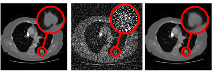
Seeing through noise: data-driven reconstruction for a simulated lung CT screening extremely low dose. Left: clinical dose, Middle: approximately 5% of clinical dose, Right: self-supervised machine learning reconstruction (noise2inverse)
Keep Reading
View all case studiesSee maths in action







Connect with us
Explore how CHIMiRA can transform your research or business challenges through mathematical innovation. We are an open house for ideas and consider projects from any academic discipline, business, industry or the third sector.
Contact us